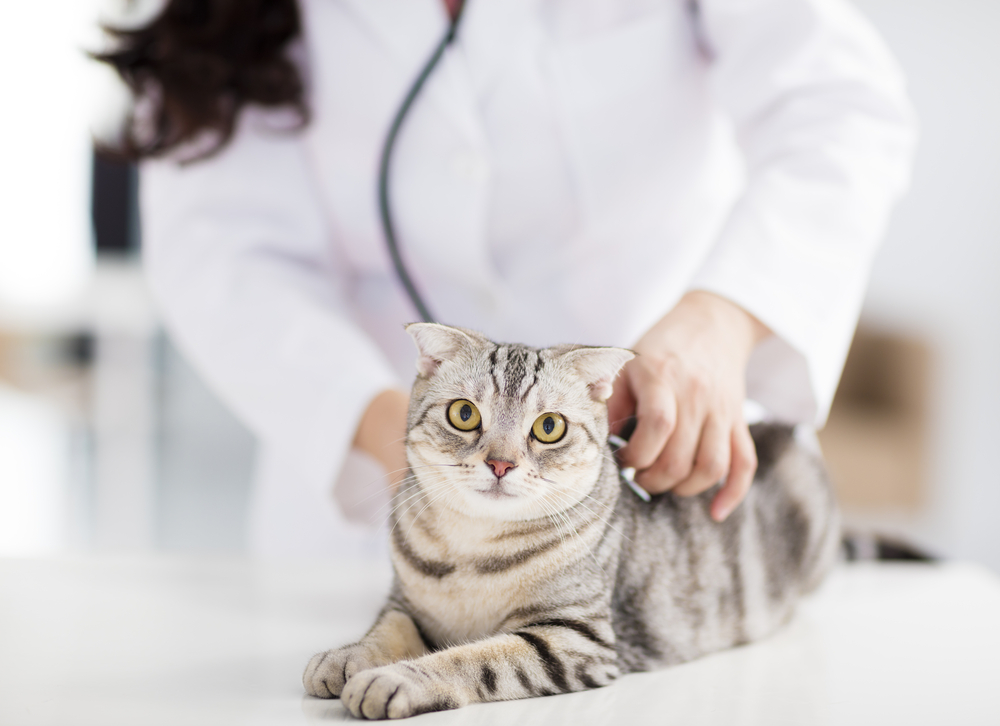
The cardiologist will perform a thorough cardiac physical exam. An important part of the cardiac evaluation is also reviewing your concerns and any signs your pet is having at home, in addition to reviewing medical records from your primary veterinarian. This information all helps to determine the best plan for your pet.
Echocardiogram
An echocardiogram is an ultrasound of the heart. This is a non-invasive test that enables us to visualize the structure, size, and function of the heart, and blood flow in and out of the heart/lungs in order to evaluate for various conditions: cardiomyopathy (disease of the heart muscle), various congenital heart diseases (diseases that animals are born with), age-related valve disease, pulmonary hypertension (elevated pressure in the pulmonary arteries/blood vessels of the lungs), and others.
Electrocardiogram
An electrocardiogram (ECG or EKG) is a non-invasive test that shows the heart rhythm, which is the electrical activity of the heart. We look for arrhythmias (abnormal heart rhythms), causing the heart to beat too slowly, too fast, or irregularly. Many arrhythmias can often be managed with medications.
Pre-Anesthesia Evaluation
Your vet may request a cardiac evaluation prior to anesthesia if a heart murmur or arrhythmia is heard on physical exam, if your pet is of a specific breed, or if your pet is having any signs that can occur with heart disease. Since heart disease can potentially increase risk for anesthesia, it is important to assess this prior, in order to determine the best course of action for your pet.
Thoracic Radiograph Interpretation
A thoracic radiograph, or chest x-ray, is a diagnostic test to evaluate the lungs and other structures in the chest. This is often recommended in pets that have an increased breathing rate or cough. Animals with advanced heart disease can develop fluid in the lungs. However, there are also various non-cardiac diseases that can cause similar signs. Thoracic radiographs can help diagnose these so an appropriate treatment plan can be started.
Holter Monitor
A Holter monitor is a device that records an electrocardiogram (ECG/EKG) over a period of 24 hours or longer. Arrhythmias (abnormal heart rhythms) can be intermittent. This means that we may not be able to see all arrhythmias on an ECG that we do in the hospital. If we suspect an arrhythmia (based on your pet’s signs or breed), we can place a Holter monitor that your pet will wear at home so that we can assess the ECG for a longer period of time.
Therapeutic Procedures
Heart disease can cause fluid to form within the lungs, around the lungs, around the heart, or in the abdomen. Fluid formation in or around the lungs or around the heart can cause trouble breathing, which is an emergency.
The treatment plan will depend on the specific diagnosis and needs of an individual pet. Heart disease may be managed with medications that you would give at home, injections given in hospital, and/or various procedures done by the cardiologist if needed, such as thoracocentesis (removal of fluid from around the lungs), pericardiocentesis (removal of fluid from around the heart), or abdominocentesis (removal of fluid from the abdomen). These procedures can be life-saving, and are sometimes necessary to provide rapid relief and improvement in breathing.
Signs of heart disease in cats
Trouble breathing: breathing rate may be faster, breaths may appear heavier, deeper, or more exaggerated. Some cats with trouble breathing may breathe at a normal rate, but each breath appears to be a deep sigh.
Restlessness (accompanied by change in breathing) – inability to sleep or rest comfortably
Collapse or fainting
Exercise intolerance: In a cat this can be transient heavier breathing after exertion, such as playing or jumping up on furniture, or stopping to lie down after taking a few steps
Cough is rarely a sign of heart disease in cats (it is more commonly due to asthma or other respiratory diseases), however some cats can cough with heart disease
Sudden paralysis or inability to use hind limbs or a forelimb
Behavior change: Although this is not specific for heart disease (it can be a sign of any other illness in a cat), when cats do not feel well they may hide or sleep in unusual places, not greet you at the normal times, or be less affectionate. Since cats are very good at hiding how they feel, it is important to have a cat evaluated when they are not acting like themselves.
Please note:
Some of these signs can also occur with other disease processes. A cardiac evaluation can determine if these signs are heart-related or not.
Cats can also have heart disease without having any obvious signs at all.
Trouble breathing is an emergency; cats with trouble breathing should go to their nearest emergency hospital.
Signs of heart disease in dogs
Trouble breathing: breathing rate may be faster, breaths may appear heavier, deeper, or more exaggerated
Cough: new cough, increase in frequency of cough, or change in sound of cough
Collapse or fainting
Exercise intolerance: getting winded or out of breath with normal activity, or not tolerating normal walks
Restlessness (accompanied by change in breathing): inability to sleep or rest comfortably
Please note:
Some of these signs can also occur with other disease processes. A cardiac evaluation can determine if these signs are heart-related or not.
Dogs can also have heart disease without having any obvious signs at all.
Trouble breathing is an emergency; dogs with trouble breathing should go to their nearest emergency hospital.